
More Information
Submitted: June 08, 2024 | Approved: June 24, 2024 | Published: June 25, 2024
How to cite this article: Bekaye T, Djénéba K, Abdoulaye K, Aissata S, Binta G, et al. Severe Atopic Dermatitis Treated by Wet Wrapping: An Observation at the Dermatology Hospital of Bamako (Mali). Clin J Nurs Care Pract. 2024; 8: 040-043.
DOI: 10.29328/journal.cjncp.1001056
Copyright License: © 2024 Bekaye T, et al. This is an open access article distributed under the Creative Commons Attribution License, which permits unrestricted use, distribution, and reproduction in any medium, provided the original work is properly cited.
Keywords: Severe atopic dermatitis; Wet wrapping; Bamako

Severe Atopic Dermatitis Treated by Wet Wrapping: An Observation at the Dermatology Hospital of Bamako (Mali)
Traoré Bekaye1*, Koné Djénéba1, Kanouté Abdoulaye1, Samaké Aissata1,2, Guido Binta1,2, Cissé Lamissa3,3, Simpara Bakary1, Diakité Mamoudou1, Dicko Adama Aguissa1,2 and Faye Ousmane1,2
1Bamako Dermatological Hospital (HDB), Mali
2Bamako University of Science, Technology and Engineering (USTTB), Mali
3Koulikoro Regional Hospital, Mali
*Address for Correspondence: Traoré Bekaye, Bamako Dermatological Hospital (HDB), Mali, Email: [email protected], [email protected]
Introduction: Wet wrapping is a local care technique adapted to the treatment of severe forms of eczema. It is a good alternative for AD resistant to the usual local treatments. We report a case.
Observation: A 5-month-old infant, with a personal history of allergic rhinitis has been seen in a dermatological for diffuse skin eruption and pruritus evolving in flare-ups since 4 months, without improvement after several courses of dermocorticoids, anti-H1, and emollient from several doctors. Clinical examination revealed erythematous plaques surmounted by vesicles with a crumbled border located on the convexities and extension face of the limbs and in the folds behind the ears and diffuse skin xerosis. The examination of the other devices was unremarkable. The evaluated SCORAD was 59.8. We carried out the treatment by the wet wrapping technique, a clear regression of the cutaneous lesions and pruritus with the decrease of the SCORAD from 59.8 to 8.8 in 1 month of treatment.
Discussion: This observation further illustrates the effectiveness of the Wet wrapping technique in the management of recalcitrant atopic dermatitis. In resource-poor countries, Wet wrapping may be an alternative for recalcitrant forms of atopic dermatitis. For fostering critical nurse observation as a source of research topics, we propose four strategies. First, cultivating awareness through a culture of evidence-based practice and critical reflection on common practice. Second, stimulating persistence in addressing moral dilemmas concerning better care despite resistance. Third, facilitating interprofessional learning in an open culture, where diverse perspectives are valued, and it is psychologically safe to bring them in. Fourth, overcoming funding disparities and facilitating nurse-led research, acknowledging the underrepresentation of nurses in funding agencies.
These measures aim to empower nurses to observe critically, use their unique perspectives, and bring in research topics.
Atopic dermatitis (AD) is a chronic, pruritic inflammatory dermatosis of genetic predisposition evolving by recurrent outbreaks [1,2].
In children, management includes, in addition to therapeutic education, emollients as background treatment, dermocorticoids for the treatment of flare-ups, and immunosuppressants for resistant and severe forms [3,4].
Wet wrapping is a local care technique adapted to the treatment of severe forms of eczema to rapidly reduce inflammation and pruritus. This technique is a good alternative for AD resistant to the usual local treatments [5]. We report a case.
A 5-month-old infant, full-term pregnancy with normal delivery, both parents are doctors, with a personal history of allergic rhinitis and more than 4 episodes of eczema and family history of eczema (father and grandmother).
He has been seen in a dermatological consultation for diffuse erythematous patches and insomniac pruritus.
The interrogation reports that these lesions have been evolving in flare-ups since the first months of life, without regression for 2 months. He received several courses of dermocorticoids (Betamethasone cream), anti-H1 (Desloratadine), and emollients from several doctors without satisfactory improvement.
Clinical examination revealed an apyretic child with more or less limited erythematous plaques surmounted by vesicles with a crumbled border located on the convexities and extension face of the limbs and in the folds behind the ears (Pictures 1,2). To the touch the lesions were rough. These lesions were visible on the convex areas (head, external surfaces of forearms, and legs). There was also a diffuse xerosis on the whole integument. The examination of the other devices was unremarkable. The evaluated SCORAD (SCORing Atopic Dermatitis) with the formula: A/5 + 7 B/2 +C, A is defined as the extent (0-100) and B is defined as the intensity (0-18) and is defined as the subjective symptoms (0-20): 47/5 + 7x9/2 + 19= 59.8 [6,7].
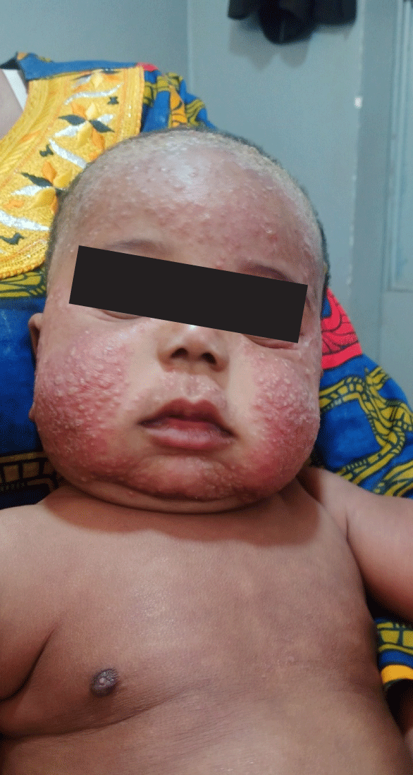
Picture 1:
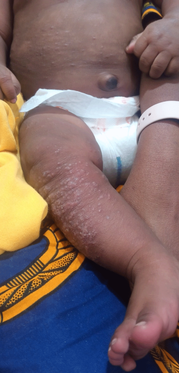
Picture 2:
We carried out the treatment by the wet wrapping technique (Picture 3), body washing with a supergras soap, application of dermocorticoid on the inflammatory zones at the rate of a phalanx for a body surface of 2 apples of hands (2,5 g for 1% body surface affected), compresses moistened with warm water and deposited on the zones to be treated then dry bandage, a daily session during 2 weeks and twice a week the remainder of the month.
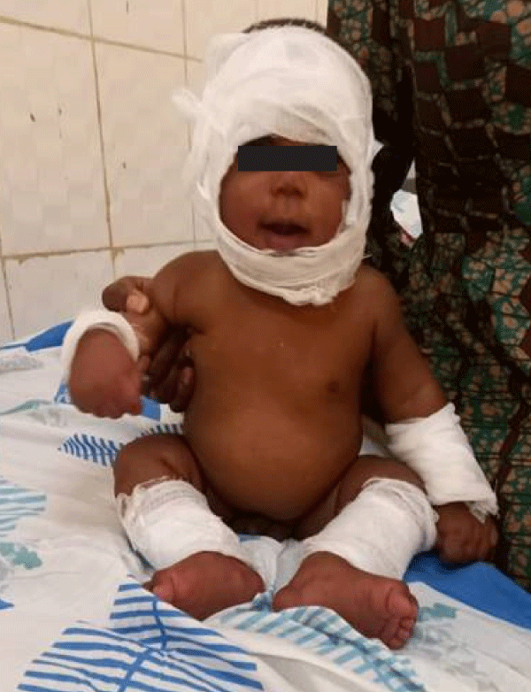
Picture 3:
The evolution was marked by a clear regression of the cutaneous lesions and pruritus with the decrease of the SCORAD from 59.8 to 8.8 in 1 month of treatment (Pictures 4,5).

Picture 4:

Picture 5:
Atopic dermatitis (AD) or atopic eczema, is a chronic inflammatory skin disease characterized by dry skin, itching, and recurrent red and scaly skin lesions [8,9]. It is a relatively common skin disease with an estimated prevalence of 10% - 20% [1,2,10]. The majority of patients show their first clinical symptoms in infancy or early childhood. The pathogenesis of AD is characterized by a complex interaction between genetic background and different environmental factors [9,11].
The diagnosis is based on clinical criteria, criteria formulated by the UK working party on AD, which have been extensively validated in the past and are widely accepted as a diagnostic tool, pruritus follows at least 3 other criteria such as skin dryness, history of asthma or allergic rhinitis and eczema beginning before 2 years old, all these criteria were found at our patient [3,12,13].
The treatment of AD is aimed at restoring the epidermal barrier defect and reducing skin inflammation, several educational programs are an effective addition to the treatment of patients with AD, reducing the severity of skin disease and improving quality of life [4].
Patients with severe and/or refractory AD may need a different therapeutic approach. Well-known intervention treatments include systemic corticosteroids, cyclosporine A, azathioprine, or photo (chemo) therapy. All of these interventions have (relative) contraindications and potential side effects, limiting their use in children [8].
Wet wrapping treatment with diluted topical corticosteroids has been advocated as a relatively safe and effective intervention treatment in children with severe and/or refractory AD [6,14-16].
Some recent data in the literature have demonstrated the effectiveness of wet wrapping to the usual treatment with dermocorticoids and emollients but the difference was not statistically significant [16,17].
Our observation further illustrates the effectiveness of the Wet wrapping technique in the management of recalcitrant atopic dermatitis. In recent years, wet wrapping treatment has been considered a safe and effective technique for severe childhood dermatitis [5]. This therapeutic modality has also been successfully used in other indications, such as erythrodermic psoriasis, treatment-resistant pruritus, active urticaria, lamellar ichthyosis, and T-cell lymphoma [15,18].
This technique protects the skin from scratching reduces pruritus and, through vasoconstriction secondary to cooling of the skin due to evaporation of moisture, reduces inflammation [4,19].
In addition, the absorption of topical corticosteroids is increased. Healing is further enhanced by the moist environment protected from external allergens.
All this leads to a marked clinical improvement, evidenced by a significant reduction in the SCORAD index.
In resource-poor countries, access to state-of-the-art molecules for the management of atopic dermatitis is exceptional. Wet wrapping may be an alternative for recalcitrant forms of atopic dermatitis.
- Bylund S, Kobyletzki LB, Svalstedt M, Svensson Å. Prevalence and Incidence of Atopic Dermatitis: A Systematic Review. Acta Derm Venereol. 2020 Jun 9;100(12):adv00160. doi: 10.2340/00015555-3510. PMID: 32412646; PMCID: PMC9189744.
- DaVeiga SP. Epidemiology of atopic dermatitis: a review. Allergy Asthma Proc. 2012 May-Jun;33(3):227-34. doi: 10.2500/aap.2012.33.3569. PMID: 22584191.
- Reynolds M, Gorelick J, Bruno M. Atopic Dermatitis: A Review of Current Diagnostic Criteria and a Proposed Update to Management. J Drugs Dermatol. 2020 Mar 1;19(3):244-248. PMID: 32550689.
- Ring J, Alomar A, Bieber T, Deleuran M, Fink-Wagner A, Gelmetti C, Gieler U, Lipozencic J, Luger T, Oranje AP, Schäfer T, Schwennesen T, Seidenari S, Simon D, Ständer S, Stingl G, Szalai S, Szepietowski JC, Taïeb A, Werfel T, Wollenberg A, Darsow U; European Dermatology Forum (EDF); European Academy of Dermatology and Venereology (EADV); European Federation of Allergy (EFA); European Task Force on Atopic Dermatitis (ETFAD); European Society of Pediatric Dermatology (ESPD); Global Allergy and Asthma European Network (GA2LEN). Guidelines for treatment of atopic eczema (atopic dermatitis) part I. J Eur Acad Dermatol Venereol. 2012 Aug;26(8):1045-60. doi: 10.1111/j.1468-3083.2012.04635.x. PMID: 22805051.
- Devillers AC, Oranje AP. Efficacy and safety of 'wet-wrap' dressings as an intervention treatment in children with severe and/or refractory atopic dermatitis: a critical review of the literature. Br J Dermatol. 2006 Apr;154(4):579-85. doi: 10.1111/j.1365-2133.2006.07157.x. PMID: 16536797.
- Vourc'h-Jourdain M, Barbarot S, Taieb A, Diepgen T, Ambonati M, Durosier V, Sibaud V, Stalder JF. Patient-oriented SCORAD: a self-assessment score in atopic dermatitis. A preliminary feasibility study. Dermatology. 2009;218(3):246-51. doi: 10.1159/000193997. Epub 2009 Jan 16. PMID: 19147989.
- Oranje AP, Glazenburg EJ, Wolkerstorfer A, de Waard-van der Spek FB. Practical issues on interpretation of scoring atopic dermatitis: the SCORAD index, objective SCORAD and the three-item severity score. Br J Dermatol. 2007 Oct;157(4):645-8. doi: 10.1111/j.1365-2133.2007.08112.x. Epub 2007 Aug 21. PMID: 17714568.
- Thomsen SF. Atopic dermatitis: natural history, diagnosis, and treatment. ISRN Allergy. 2014 Apr 2;2014:354250. doi: 10.1155/2014/354250. PMID: 25006501; PMCID: PMC4004110.
- Li H, Zhang Z, Zhang H, Guo Y, Yao Z. Update on the Pathogenesis and Therapy of Atopic Dermatitis. Clin Rev Allergy Immunol. 2021 Dec;61(3):324-338. doi: 10.1007/s12016-021-08880-3. Epub 2021 Aug 2. PMID: 34338977.
- Lee HH, Patel KR, Singam V, Rastogi S, Silverberg JI. A systematic review and meta-analysis of the prevalence and phenotype of adult-onset atopic dermatitis. J Am Acad Dermatol. 2019 Jun;80(6):1526-1532.e7. doi: 10.1016/j.jaad.2018.05.1241. Epub 2018 Jun 2. PMID: 29864464.
- Peng W, Novak N. Pathogenesis of atopic dermatitis. Clin Exp Allergy. 2015 Mar;45(3):566-74. doi: 10.1111/cea.12495. PMID: 25610977.
- Brenninkmeijer EE, Schram ME, Leeflang MM, Bos JD, Spuls PI. Diagnostic criteria for atopic dermatitis: a systematic review. Br J Dermatol. 2008 Apr;158(4):754-65. doi: 10.1111/j.1365-2133.2007.08412.x. Epub 2008 Jan 30. PMID: 18241277.
- Williams HC, Burney PG, Pembroke AC, Hay RJ. The U.K. Working Party's Diagnostic Criteria for Atopic Dermatitis. III. Independent hospital validation. Br J Dermatol. 1994 Sep;131(3):406-16. doi: 10.1111/j.1365-2133.1994.tb08532.x. PMID: 7918017.
- Oranje AP, Devillers AC, Kunz B, Jones SL, DeRaeve L, Van Gysel D, de Waard-van der Spek FB, Grimalt R, Torrelo A, Stevens J, Harper J. Treatment of patients with atopic dermatitis using wet-wrap dressings with diluted steroids and/or emollients. An expert panel's opinion and review of the literature. J Eur Acad Dermatol Venereol. 2006 Nov;20(10):1277-86. doi: 10.1111/j.1468-3083.2006.01790.x. PMID: 17062046.
- Bingham LG, Noble JW, Davis MD. Wet dressings used with topical corticosteroids for pruritic dermatoses: A retrospective study. J Am Acad Dermatol. 2009 May;60(5):792-800. doi: 10.1016/j.jaad.2008.12.043. PMID: 19389521.
- Dabade TS, Davis DM, Wetter DA, Hand JL, McEvoy MT, Pittelkow MR, el-Azhary RA, Davis MD. Wet dressing therapy in conjunction with topical corticosteroids is effective for rapid control of severe pediatric atopic dermatitis: experience with 218 patients over 30 years at Mayo Clinic. J Am Acad Dermatol. 2012 Jul;67(1):100-6. doi: 10.1016/j.jaad.2011.06.025. Epub 2011 Oct 5. PMID: 21978575.
- Hindley D, Galloway G, Murray J, Gardener L. A randomised study of "wet wraps" versus conventional treatment for atopic eczema. Arch Dis Child. 2006 Feb;91(2):164-8. doi: 10.1136/adc.2004.050831. Epub 2005 Nov 24. PMID: 16308411; PMCID: PMC2082699.
- Andersen RM, Thyssen JP, Maibach HI. The role of wet wrap therapy in skin disorders – a literature review. Acta Derm Venereol. 2015 Nov;95(8):933-9. doi: 10.2340/00015555-2134. PMID: 25940919.
- Devillers AC, Oranje AP. Wet-wrap treatment in children with atopic dermatitis: a practical guideline. Pediatr Dermatol. 2012 Jan-Feb;29(1):24-7. doi: 10.1111/j.1525-1470.2011.01691.x. PMID: 22256990.